Pediatric Transplant Research Laboratory

Two New Approaches Seek to Improve the Effectiveness of Anti-rejection Drugs

The Pediatric Transplant Research Laboratory, directed by Rakesh Sindhi, MD, is developing ways to improve the effectiveness of anti-rejection medicines, such that each child can receive the medicine most appropriate for her, at a dose that prevents rejection, but does not cause side effects.

Genomic fingerprints

Two approaches are being developed. The first approach has identified preliminary genomic fingerprints that differentiate children prone to rejection, from those that never experience rejection, on the usual anti-rejection medicines. These “fingerprints” are based on mutations, also known as “single nucleotide polymorphisms”, in the “immune response” area of the genome called the major histocompatibility complex (The photographer took a picture from a computer screen that represents this area). These fingerprints will someday allow transplant physicians to select the right anti-rejection medicine for a given child before transplantation.

Identifying the risk of transplant rejection

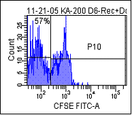
The second approach is to identify after transplantation, the risk of rejection at any given time, especially when reduction in anti-rejection medication doses are being contemplated. This is done by measuring the extent to which white blood cells from recipients proliferate, or divide into daughter cells, upon coming in contact with similar cells from their donor, or from an unrelated human being (third-party). Rejection is often described as the selective response of a recipient toward a donor. The figure shown below is from an individual who is in the midst of a liver rejection event. Proliferating cells shown in the left hand area (57%) in response to donor, greatly exceed proliferating cells in response to third-party (10%).

This figure is from an individual who is in the midst of a liver rejection event.    This image is from an individual who is in the midst of a liver rejection event.

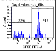
In the case of another child in whom rejection was not present at the time of biopsy (see figure below), cells proliferating in response to donor (33%) were far less than those proliferating in response to
third-party (45%).

In the case of another child in whom rejection was not present at the time of biopsy cells proliferating in response to donor.  In the case of another child in whom rejection was not present at the time of biopsy cells proliferating in response to donor.

Learn more about Quality of Life Improvements for Organ Transplant Recipients.